Study of Worker Bee Morphology
Title :- Study of Worker bee morphology
External Morphology of Worker bee :-
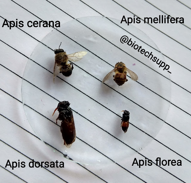
The worker bee is smallest member of the colony. It is black brown in colour and entire body is densely covered with hairs. The body of worker bee is divided into three regions, Head, Thorax and Abdomen.
• Mouth Parts :- These are attached to the lower part of head. The mouth parts are Biting and Sucking type. It consists of labrum, epipharynx, mandibles, maxillae and labium.
1)Labrum - Large plate like attached to lower margin of clypeus.
2)Epipharynx - It lies below the labrum. Fleshy in appearance. Epipharynx is a organ of taste.
3)Mandibles - These are two in number and lies on the sides of labrum. The mandibles of worker are spoon shaped, thick at the base and narrowed through the middle. At the base of mandibles, mandibular glands open. Mandibles are equipped with abductor and adductor muscles which work sidewise. Mandibles are useful to gather Pollen and mould the wax.
4)Maxillae - It lies beneath mandibles. The lacinia is absent in maxillae while the maxillary palps are vestigial and the galea is elongated and blade-like.
5)Labium - The labium shows strongly reduced Paraglossae but the glossae are very much elongated. They are united, hairy and form a honey-spoon called labellum at the terminal part. The labial palps are well developed and help to make the ligula up. The apparatus is well surrounded by galeae of maxillae. At the time of nectar feeding, the labium and maxillae come together to form sucking tube.
• Wings :- The wings are small, narrow, membranous and transparent. They lie flat over the back at rest. Wings shows modified and reduced wing veination. The fore and hind wings are interlocked by hooks (hamuli), so as to work together during flight.
• Legs :- There are three pairs of legs, i.e. prothoracic, mesothoracic & metathoracic shows progressive increase in length from 1st to 3rd pair. Legs are densely covered with hairs. Each leg consists of five parts viz., coxa, trochanter, femur, tibia and tarsus. The tarsus is five joined and ends into the claws and pulvillus. Each pair of legs are structurally modified to suit various activities / functions.
a)Prothoracic Leg - Number of stiff bristles are present on the anterior face of tibia distally, which forms Pollen brush. On the posterior face of tibia have movable plate like process called velum, which fits over a circular notch in the upper part of the first tarsal segment. The velum and antena- comb together serve as antenna cleaners.
Eye brush is present on the anterior surface of first tarsal segment which is used for removing pollen and other particles from the surface of compound eyes.
b)Mesothoracic Leg - The middle leg shows the usual portion. The inner surface of the tibial segment bears a brush and a spine-like (pollen spur) at its distal end. The spurs are used to remove pollen from the pollen baskets of the hind legs and to remove wax from the wax pockets on the ventral surface of the abdomen.
c)Metathoracic Leg - Each metathoracic leg consists of a large tibia which contains a cavity and forms the pollen basket. i.e. a depression on the outer surface of tibia, used for storing pollens during collection. At the distal end the tibia has a row of stiff bristles called pectins below which has a flat plate, known as auricle. The pectines and auricle form a pollen packet to convey packed pollens into the Pollen basket.
• Abdomen :- The abdomen of worker bee is oval and posterior most region of the body. It has six visible segments.
i.e. II to VII because the first segment (propodeum) transferred to thorax and remaining are reduced. Each visible segment has large dorsal tergum, and smaller ventral sternum. The successive terga and sterna are connected by intersegmental membrane. The posterior part of abdomen is modified into sting apparatus and wax gland on ventral surface of abdomen.
• Sting apparatus :- In the worker bee, the ovipositor changes so as to take the function of sting i.e. for injecting poison. The sting apparatus or poison apparatus has three components, namely, the sting, the plates and poison gland.
1)The Sting - The sting is a hollow organ formed by three pieces connected to a central canal (venom/poison canal). The dorsal part is the stylet sheath and the two ventral pieces are stylets or lancets. The tips of the stylets and their sheaths have forward-directed barbs. The stylet, the sheath widening at its base into the bulb of the sting and then into a pair of twisted arms.
2)The plates - Three Pairs of plates associated with the sting act as a lever. The innermost pair of oblong plates posterior in position and representing the divided 9th sternum. Two triangular or Fulcral plates representing reduced 8th sternum and attached to corresponding stylets. The large quadrate plates lie dorsally to the triangular plates and at the posterior angle.
3)Poison gland -
There are two glands -
i)Acid glands - It is elongated, slender gland open into the upper end of poison sac. It discharges an acidic secretion into sac.
ii)Alkaline gland - It is thick, tubular gland which opens externally below the base of the bulb.
-----------------THANK YOU-----------------


Post a Comment